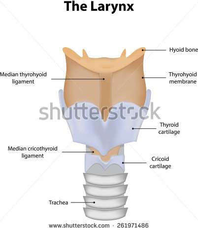
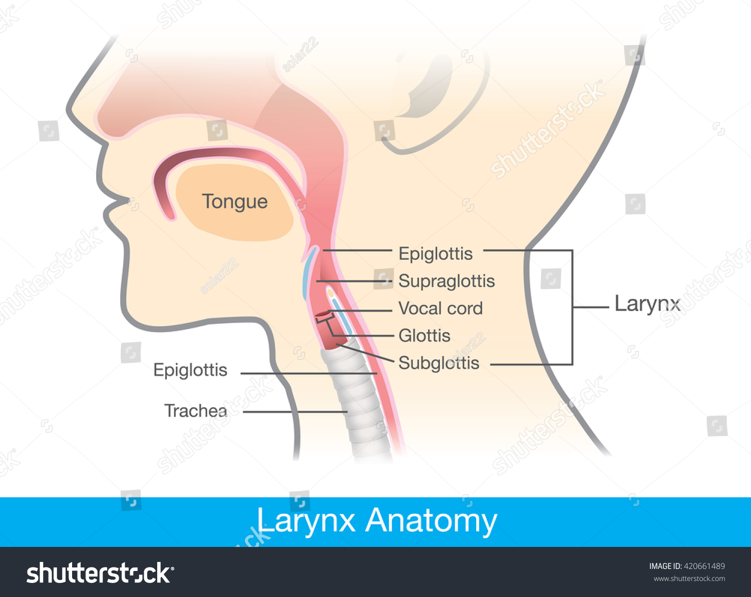
40 larynx diagram labeled
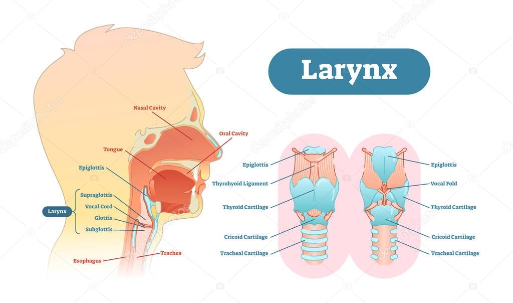
The larynx (/ ˈ l æ r ɪ ŋ k s /), commonly called the voice box, is an organ in the top of the neck involved in breathing, producing sound and protecting the trachea against food aspiration. The opening of larynx into pharynx known as the laryngeal inlet is about 4-5 centimeters in diameter. The larynx houses the vocal cords, and manipulates pitch and volume, which is essential for ... Muscles of the larynx. There are many muscles that either make up a certain part of the laryngeal structure inside the neck, or that sit adjacent to it and aid in its function.These muscles produce the movements of the larynx and its cartilages, thus enabling the proper air conduction, speech, movements of the epiglottis and airways protection. The muscles of the larynx are divided into two ...
Browse 1,594 larynx stock photos and images available, or search for larynx anatomy or larynx surgery to find more great stock photos and pictures. An anatomical diagram consisting of a vertical cross-section of the human head, and showing the relations of the nasal and buccal cavities and the...
Larynx diagram labeled
Anatomy and Physiology 2 Laboratory Manual. Main Body. Fetal Pig Dissection. ... Below the oropharynx, the laryngopharynx leads to the opening of the larynx and esophagus. From the laryngopharynx, air passes through the glottis to the trachea. Below: hard palate, soft palate, glottis, epiglottis, tongue ... The diagrams below summarize the ... This laryngeal poster digital download duo print set is a pretty and functional reminder to keep in your home office/study space! Perfect for dorm rooms, home offices, workspaces, and more, this print makes a great gift for supervisors, SLPAs, SLPs, students, mentors, therapists, professors, and The Larynx. View Article. Laryngeal Cartilages. View Article. Laryngeal Ligaments and Folds. View Article. Laryngeal Muscles. View Article. Anatomy Video Lectures. START NOW FOR FREE. TeachMe Anatomy. Part of the TeachMe Series. The medical information on this site is provided as an information resource only, and is not to be used or relied on ...
Larynx diagram labeled. Larynx Diagram. Anatomy of the Larynx. It is made up of multiple pieces of tough cartilage, surrounded and held together by fibrous tissues, membranes, and ligaments [3]. The largest cartilaginous segment is called the thyroid cartilage, with a prominent bulging known as Adam’s apple. Understanding the Basics of Throat Anatomy with Diagram and Pictures. The Throat is one of the most complex parts of the human body. It starts from the pharynx and extends to the upper end of the esophagus. Check Here to Understand the Function and Part of it. Feb 11, 2018 · The Larynx Labeled Diagram - buy this illustration on Shutterstock & find other images. What is larynx (voice box) definition, where is it located, anatomy (cartilages, muscles, innervations), what does the larynx do, picture, diagram. Picture of Larynx and Vocal Cords Labeled Diagram stock photo, images and stock photography. ANATOMY OF RESPIRATORY TRACT •Anatomically Respiratory tract is divided into upper and lower tract in relation to vocal cord. Upper: nose, mouth, pharynx, larynx, trachea, and mainstem bronchi. ... the larynx at the lower border of cricoid cartilage at the level of C6, and terminates at the carina (the
Larynx anatomy with labeled structure scheme and educational medical views Larynx anatomy with labeled structure scheme and educational medical views. Anterior, posterior and cross section examination with trachea parts vector illustration. Vocal cords housing description. larynx stock illustrations Feb 11, 2018 - Explore Dana's board "Larynx model" on Pinterest. See more ideas about speech and language, speech language pathology, speech pathology. Download 1,657 Trachea Diagram Stock Illustrations, Vectors & Clipart for FREE or amazingly low rates! New users enjoy 60% OFF. 178,107,400 stock photos online. Find Larynx Vocal Cord Labeled Diagram stock images in HD and millions of other royalty-free stock photos, illustrations and vectors in the Shutterstock collection. Thousands of new, high-quality pictures added every day.
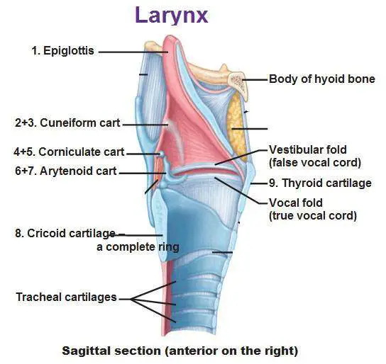
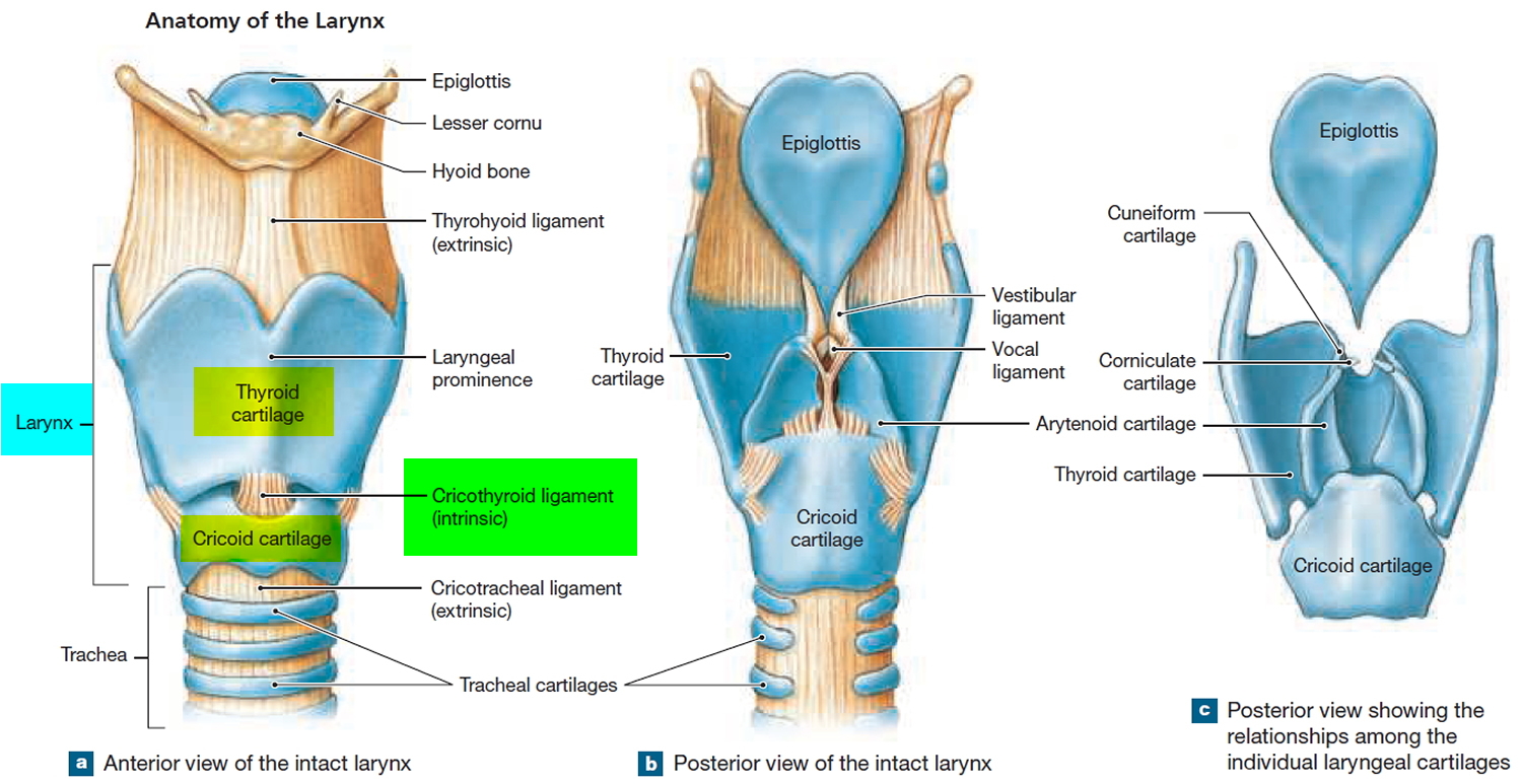
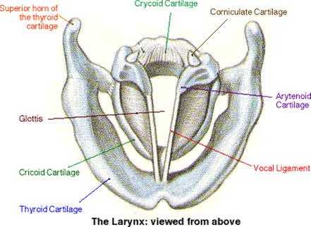
General Anatomy of the Larynx – Larynx Anatomy. The walls of the larynx are made up of cartilage, ligaments, membranes, muscles, and respiratory mucosa (or mucous membrane ). There are nine (9) laryngeal cartilages, three (3) paired and three (3) single single. Together, they form a supportive skeletal framework. Larynx anatomical vector illustration diagram, educational medical scheme. Larynx anatomical vector illustration diagram, educational medical scheme with nasal cavity, larynx, trachea and esophagus. human throat anatomy stock illustrations The larynx is a guarded air passageway between the pharynx and the trachea. It is formed by 9 supportive cartilages, intrinsic and extrinsic muscles and a mucous membrane lining. It is a short 1.5 inch tube that is located in the throat, inferior to the hyoid bone and tongue and anterior to the esophagus. Intrinsic Muscles of the Larynx. Illustration about Human larynx anatomy. this illustration is medical diagram. Illustration of normal, breathing, disease - 71512582
Anatomy Of The Larynx - Anatomy Diagram Book
Anatomy . The larynx is a system of mucosal folds supported by a cartilaginous framework. Tension and movement of the mucosal folds is effected by the actions of small muscles pulling against this cartilaginous framework. Mucosa . ... Diagram of a supraglottic laryngectomy. Dotted line shows the incision of a supraglottic laryngectomy, passing ...

Larynx anatomy | enteducationswansea
Draw a labelled diagram of larynx and explain its functions. sound class-8 1 Answer +1 vote answered Jun 25, 2020 by Renu01 (52.6k points) selected Jun 25, 2020 by BhratJha Best answer In humans, the sound is produced by the box, which is called voice box or the larynx. Larynx is a part of the throat. It is responsible for production of sound.
Pharynx (illustration) | Image | Radiopaedia.org
Best viewed on 1280 x 768 px resolution in any modern browser. Diagram of larynx 1303. Diagram of larynx 1328. Diagram of larynx 1329. Diagram of larynx 1336. Diagram of larynx 1340. Diagram of larynx 1342. Diagram of larynx 1348. Diagram of larynx 1356.

Larynx | Contemporary Health Issues
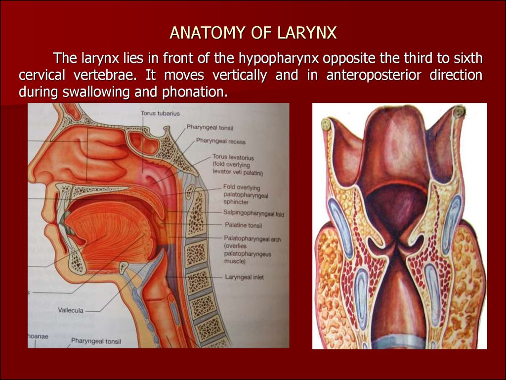
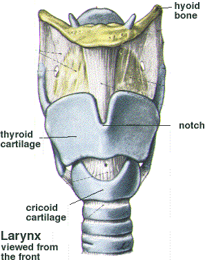
Larynx (anterior view) The larynx is a complex hollow structure located in the anterior midline region of the neck.It is anterior to the esophagus and at the level of the third to the sixth cervical vertebrae in its normal position. It consists of a cartilaginous skeleton connected by membranes, ligaments and associated muscles that suspend it from surrounding structures.

Black Rabbit, Byker Farm, Ouseburn Valley, Newcastle Upon Tyne, Tyne & Wear, England.
Vector illustration on isolated background. Cross section of the larynx above the vocal cords, with the parts, 1: right vocal cord. 2: left vocal cord. 3: cartilages to which the vocal cords are attached behind and 4: front edge of the larynx, vintage line drawing or engraving illustration. Larynx and pharynx anatomy human head anatomy ...

unknown
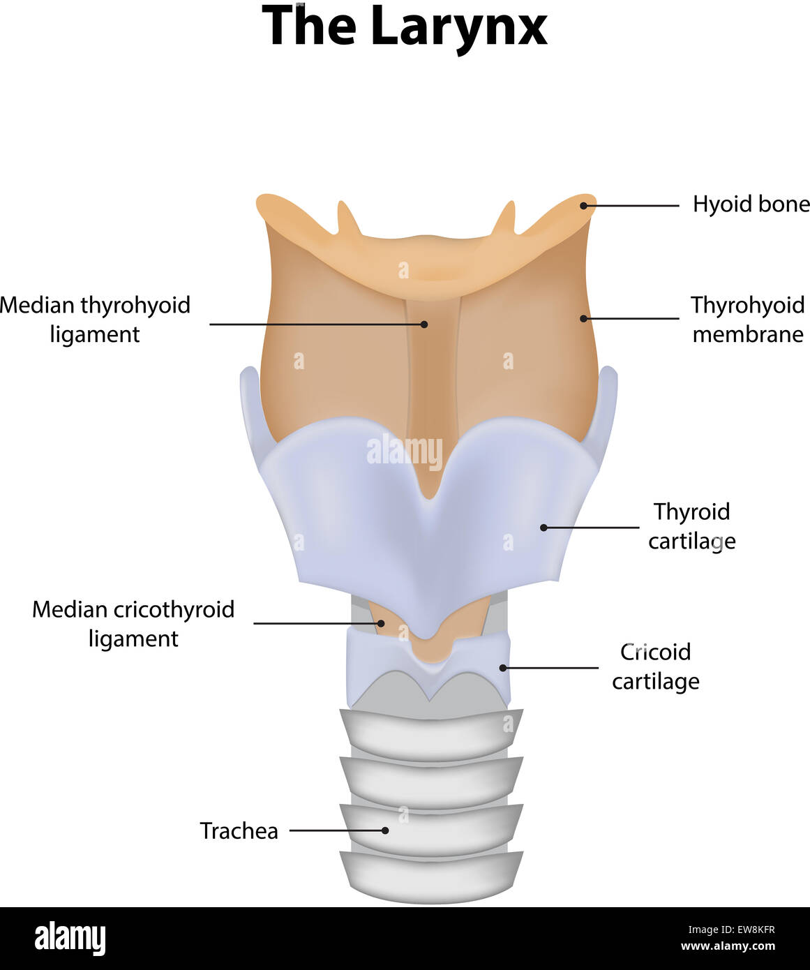
The larynx is a 4 cm long structure below an almost 2 cm inlet ( Fig. 1-6 ). It overlies the 4th, 5th, and 6th cervical vertebra in adult males, higher in females and children. It is suspended from the hyoid bone via a flexible sheet of ligament, the thyrohyoid membrane ( Fig. 1-6 ). The hyoid bone is secured dorsally to the skull via the SHL.
Larynx diagram
Larynx anatomy with labeled structure scheme and educational medical views. 1. Editable Vector .AI file. 2. Editable Vector .EPS-10 file. 3. High-resolution JPG image. Use for everything except reselling item itself. Description: Larynx anatomy with labeled structure scheme and educational medical views.
navigation map
Start studying Label The Larynx. Learn vocabulary, terms, and more with flashcards, games, and other study tools.

The Larynx
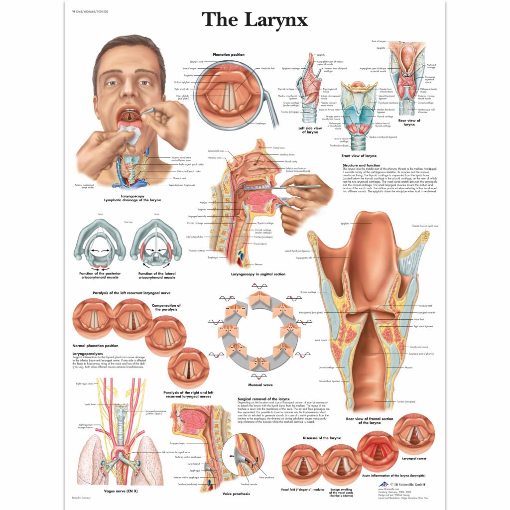
B: Schematic diagram showing the relationship of the deep spaces of the larynx within the supraglottic larynx and more superficial landmarks. FIGURE 201.8. Diagram showing the relationship of the true vocal cords to the laryngeal skeleton and various connecting membranes.
white and yellow ice cream with cone
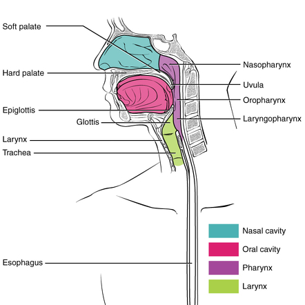
Pharynx Anatomy. Description: Anatomy of the pharynx; drawing shows the nasopharynx, oropharynx, and hypopharynx. Also shown are the nasal cavity, oral cavity, hyoid bone, larynx, esophagus, and trachea. Anatomy of the pharynx (throat). The pharynx is a hollow tube that starts behind the nose, goes down the neck, and ends at the top of the ...
Larynx Part I
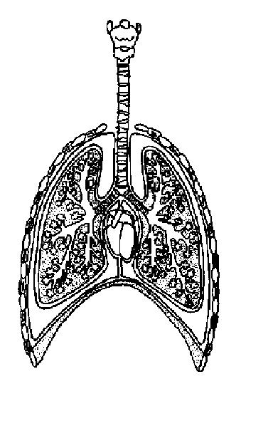
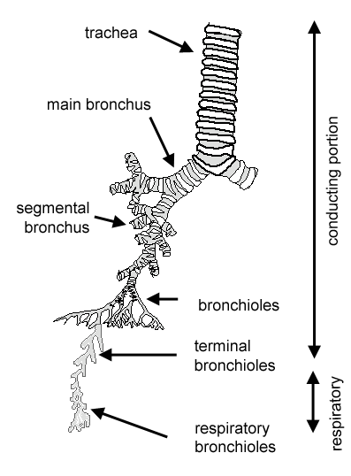
larynx. • After passing through the larynx, air enters the . trachea, which is held open by incomplete rings of cartilage. ... • Label the diagram: Page 4. Demonstration of Pleurae and the Lungs • Each lung is surrounded by two layers of. serous membrane known as the pleurae. • The relationship between the pleurae and the lungs can be ...

Do You Have a Voice Disorder? | 1SpecialPlace
Anatomy. Function. Associated Conditions. Tests. Commonly called the voice box, the larynx is located on top of the neck and is essential for breathing, vocalizing, as well as ensuring food doesn’t get stuck in the trachea and cause choking. Sitting just in front of the esophagus, the vocal folds are located here, making this organ absolutely ...
Coronal section through the larynx to show the main ...
The Larynx. View Article. Laryngeal Cartilages. View Article. Laryngeal Ligaments and Folds. View Article. Laryngeal Muscles. View Article. Anatomy Video Lectures. START NOW FOR FREE. TeachMe Anatomy. Part of the TeachMe Series. The medical information on this site is provided as an information resource only, and is not to be used or relied on ...
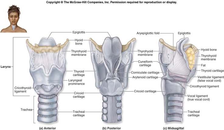
Diagram of larynx
This laryngeal poster digital download duo print set is a pretty and functional reminder to keep in your home office/study space! Perfect for dorm rooms, home offices, workspaces, and more, this print makes a great gift for supervisors, SLPAs, SLPs, students, mentors, therapists, professors, and

unknown
Anatomy and Physiology 2 Laboratory Manual. Main Body. Fetal Pig Dissection. ... Below the oropharynx, the laryngopharynx leads to the opening of the larynx and esophagus. From the laryngopharynx, air passes through the glottis to the trachea. Below: hard palate, soft palate, glottis, epiglottis, tongue ... The diagrams below summarize the ...
The Larynx Chart - SEM Trainers
Larynx - Anatomy, Function in Respiratory System - Cancer ...
Larynx anatomy

white printer paper with green line
Larynx Labeled Diagram Stock Photo - Alamy

Pin on Speech Language Pathology
Larynx anatomical vector illustration diagram, educational ...
white animal skull on white surface

Anatomy Of The Larynx - Anatomy Diagram Book
Respiratory System Worksheet - WikiEducator

Schematic representation of the sagittal anatomy of the ...

yellow green and white map

#epiglottis #anterior #posterior #median #anatomy | Throat ...
Larynx diagram | Healthiack
shallow focus photo of book page
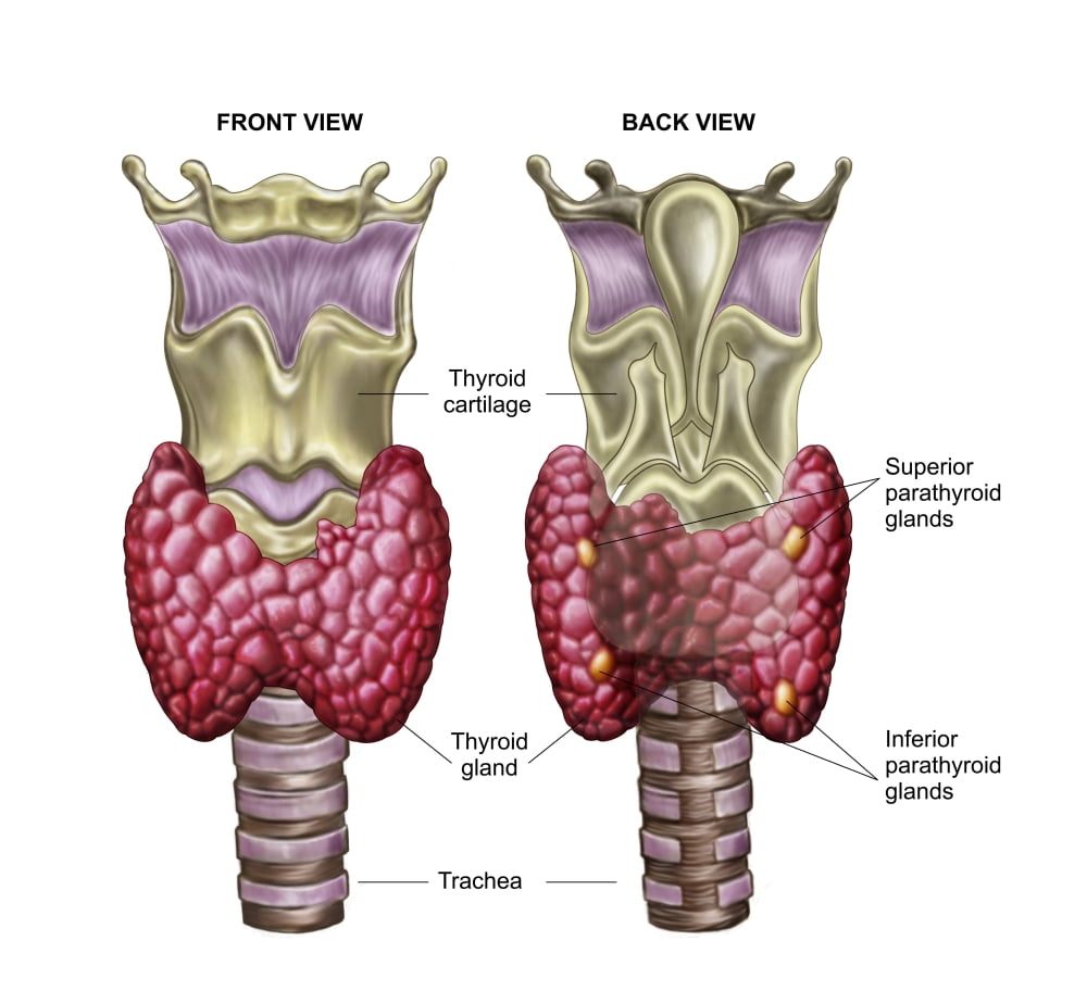
Anatomy of thyroid gland with larynx & cartilage Poster ...

Hyoid Bone Illustrations, Royalty-Free Vector Graphics ...
Human Larynx Anatomy. This Illustration Is Medical Diagram ...
![[DIAGRAM] Parts Of The Larynx Diagram FULL Version HD ...](http://www.artandsciencegraphics.com/wp-content/uploads/Larynx-Front-View.jpg)
[DIAGRAM] Parts Of The Larynx Diagram FULL Version HD ...

Schematic of the human larynx framework, based on Gray [6 ...

Pharynx & Larynx Anatomical Chart - Anatomy Models and ...
Respiratory: The Histology Guide
unknown
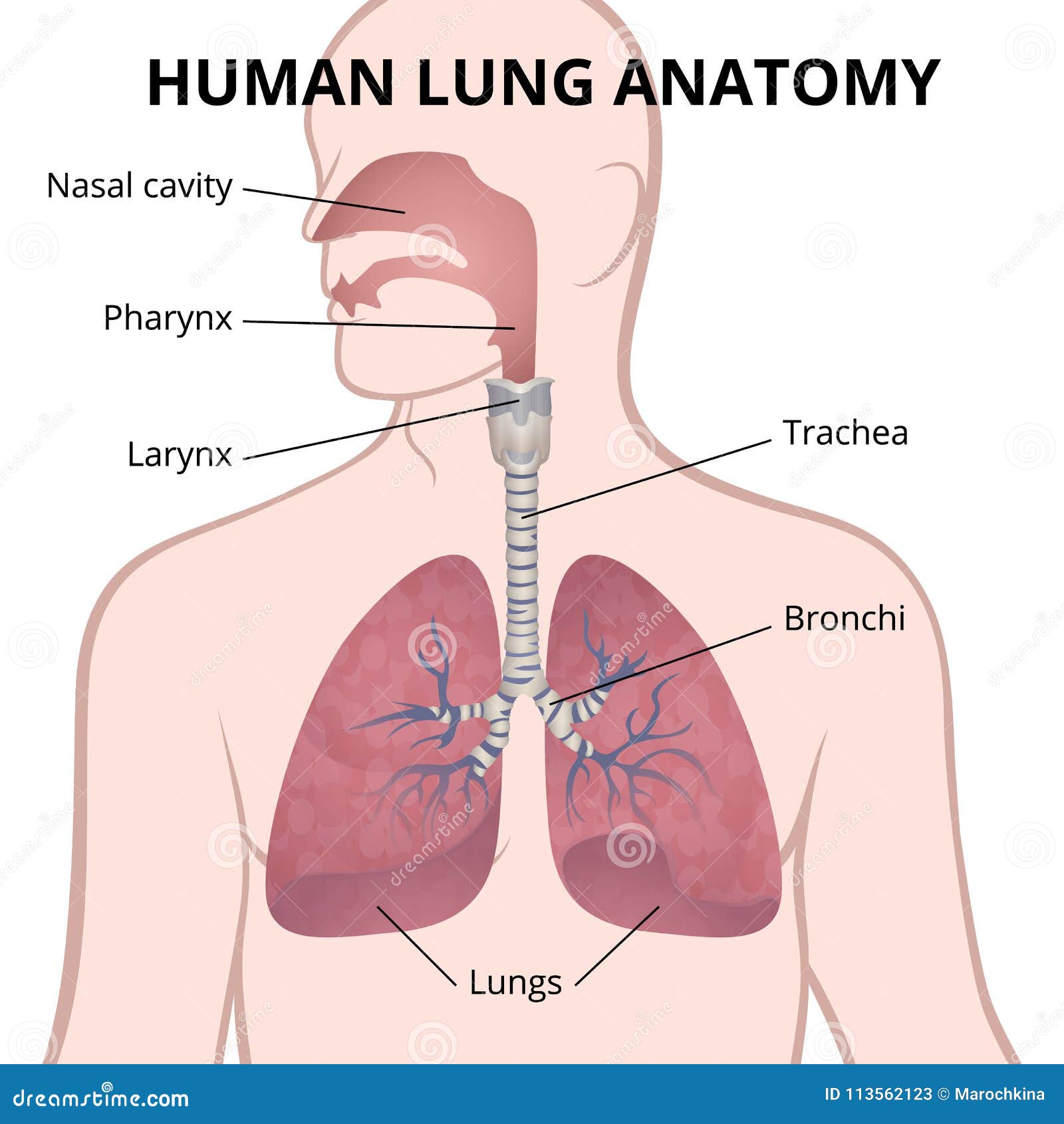
Human Lungs, Trachea And Nasopharynx Stock Vector ...

white spiral paper on black surface


Comments
Post a Comment