40 diagram and label a chromosome in prophase
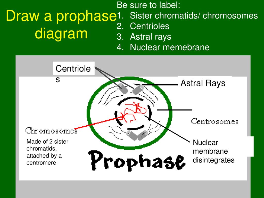
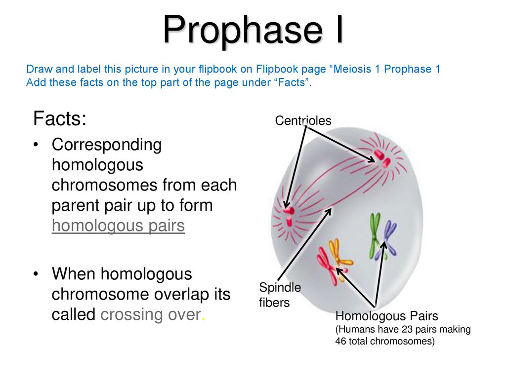
26 Diagram And Label A Chromosome In Prophase - Labels Ideas ... Mar 25, 2020 · Diagram and label a chromosome in prophase mitosis 2n 4 youtube unbelievable diagram and label a chromosome in prophase. Cytokinesis division of the cytoplasm and the formation of two distinct cells occurs. Diagrams of mitosis the process of cell division via mitosis occurs in a series of stages including prophase metaphase anaphase and telophase. Prophase Whitefish Blastula Cell The spindle fibers first appear between the separating centriole pairs. Other fibers radiate out from the centrioles to create the aster. These fibers brace the centriole and spindle apparatus against the cell membrane. Label the prophase diagram: 1. centriole pair, 2. aster, 3. spindle, 4. chromosome.
Telophase Labeled Diagram - schematron.org Nov 22, 2018 · Telophase Labeled Diagram. Prophase I: Chromosomes condense. Because of replication during interphase , each chromosome consists of 2 sister chromatids joined by a centromere. Diagrams showing the stages of mitosis including diagrams of interphase, diagrams of prophase, diagrams of metaphase, diagram of anaphase and diagram of.
Diagram and label a chromosome in prophase
Bio Chapter 10 Flashcards - Quizlet Which event occurs only during prophase I of the first meiotic division? Synapsis of homologous chromosomes occurs. Which of the following is included in alternation of generations? All of the above : Zygote, spores, multicellular diploid stage (sporophyte), multicellular haploid stage (gametophyte) What is a locus? the precise location of a gene on a chromosome. How many … Meiosis I and Meiosis II; Life Cycles - Montgomery College Prophase II The chromosomes coil up, the nuclear membrane begins to disintegrate, and the centrosomes begin moving apart. 6. Metaphase II Spindle fibers form and sister chromatids align to the equator of the cell. ... Label the diagram (1-11) with phase or cell type (for what applies to the corresponding number), and chromosome number. ... Chromosomes Fact Sheet - Genome.gov The unique structure of chromosomes keeps DNA tightly wrapped around spool-like proteins, called histones. Without such packaging, DNA molecules would be too long to fit inside cells. For example, if all of the DNA molecules in a single human cell were unwound from their histones and placed end-to-end, they would stretch 6 feet.
Diagram and label a chromosome in prophase. DOC Draw labeled diagrams showing the 5 stages of mitosis ... Draw labeled diagrams showing the 5 stages of mitosis (prophase, prometaphase, metaphase, anaphase and telophase) for a haploid cell where N=4. Use 4 different shapes to represent the different chromosomes that make up a complete set. Make sure you show the number, shape, color and location of the chromosomes at each stage of division. PDF Cell Division Animal Cell and Mitosis Key 1. Label the four phases of mitosis in the diagram. 2. Label the spindles and centrioles in one of the phases. 3. Color each chromosome in prophase a different color. Follow each of these chromosomes through mitosis. Show this by coloring the correct structures in each phase of mitosis. les Interphase Cytokinesis hase Chromatin Chromosome /(/t ... Diagram and label a chromosome in prophase Diagram and label a chromosome in prophase. Answers: 2 Show answers Another question on Biology. Biology, 21.06.2019 23:00. What are tissues? a groups of tissues that work together b basic units of structure and function c groups of cells that perform the same function d multiple organs that perform a major function ... Diagram and label a chromosome in prophase Diagram and label a chromosome in prophase. Answers: 2 Show answers Another question on Biology. Biology, 21.06.2019 23:00. Use the drop down menus to match each example to the fossil topic discussed then to show how the fossil record gives evidence of evolution ...
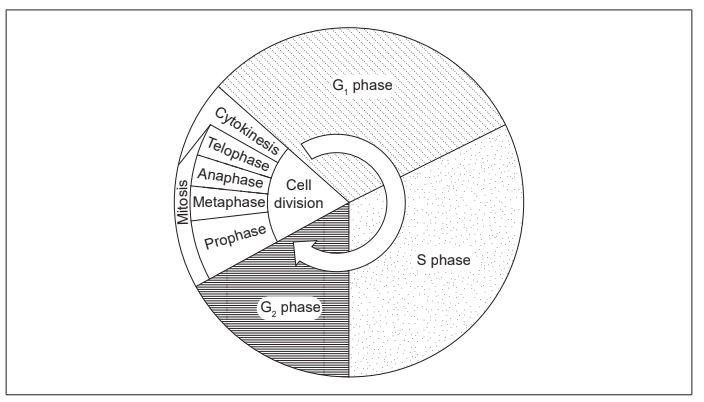
THE CELL CYCLE WORKSHEET - Chandler Unified School District The diagram below shows six cells in various phases of the cell cycle. Note the cells are not arranged in the order in which the cell cycle occurs. Use the diagram to answer questions 1-7. Cells A & F show an early and a late stage of the same phase of the cell cycle. What phase is it? prophase . Which cell is in metaphase? C Mitosis Worksheet & Diagram Identification Using the labeled Cell Cycle diagram to the right do the following: Left column: place the corresponding letter for the correct stage . Right column: Complete the description of the stage in the. A _____: Consists of G1, S and G2 phases in the Cell Cycle. ___ Cytokinesis – division of the cytoplasm resulting in two identical . d _____ cells. ___ G1 – growth phase: Makes organelles … Mitosis - Wikipedia Label-free live cell imaging of Mesenchymal Stem Cells undergoing mitosis. Onion cells in different phases of the cell cycle enlarged 800 diameters. a. non-dividing cells b. nuclei preparing for division (spireme-stage) c. dividing cells showing mitotic figures e. pair of daughter-cells shortly after division. In cell biology, mitosis (/ m aɪ ˈ t oʊ s ɪ s /) is a part of the cell cycle in ... Stages Of Meiosis Diagram Labeled - wiringall.com Simple Boat Wiring Diagram. Browning Buckmark Parts Diagram. Giant Octopus Diagram. Dec 12, · a diagram showing the various phases of meiosis such as prophase, anaphase, metaphase, telophase. each diagram will show the changes taking place in each phase, such as the arrangement of chromatids alongwith each other., or the positions of chiasma.
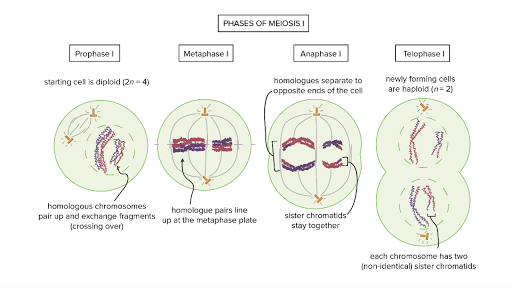
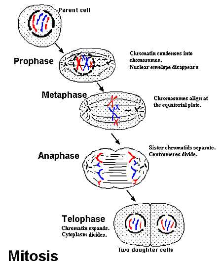
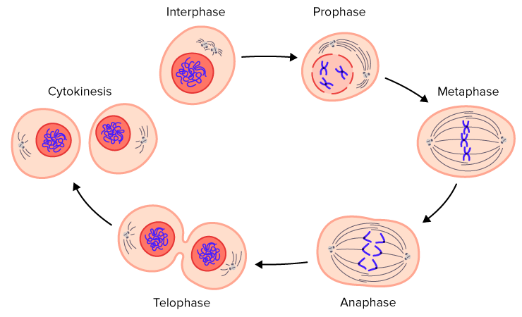
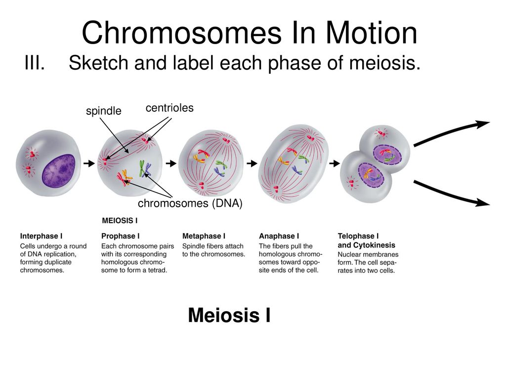
Meiosis and Mendel Study Guide A - images.pcmac.org sex chromosome 15. Mitosis 16. Meiosis 17. a; b; e SECTION 2. PROCESS OF MEIOSIS 1. sister chromatid 2. homologous 3. Refer to Figure 2.3 for visual answers. prophase I 4. metaphase I 5. anaphase I 6. telophase I 7. prophase II 8. metaphase II 9. anaphase II 10. telophase II 11. anaphase I should be circled; anaphase II should be boxed. 12. Sperm cell … Prophase Diagrams - Wiring Diagrams Free Oct 21, 2018 · Prophase, metaphase, anaphase, and telophase. Diagrams of Mitosis - the process of cell division via mitosis occurs in a series of stages including prophase, metaphase, Anaphase and Telophase. It is easy to describe the stages of mitosis in the form of diagrams showing the dividing cell (s) at each of the main stages of the process. Prophase Diagrams - schematron.org In the upper diagram two tetrads are represented as two x-shaped chromosomes associated side by side. Interphase Prophase Prometaphase The cell prepares for mitosis. At this stage, the chromosomes are not clearly visible. The chromosomes, pairs of sister chromatids, condense and become visible. Prophase - Yale University Prophase At prophase the replicated chromosomes condense. Compare the staining of the chromosomes in the prophase cell to those in the surrounding, non-dividing cells. Each chromosome in prophase consists of two sister chromatids. Note that the nuclear envelope is still intact.
Label the diagram below. Use these choices: Metaphase 1 ... Prophase 1: First stage of Meiosis 1.The centrioles move to the poles of the cell, the nuclear membrane disintegrates, homologous chromosomes pair up (in the form of tetrad), form a chiasmata and then exchange segments of chromosomes with each other. This process is called crossing over. Metaphase 1:
Prophase Animal Cell Diagram - Studying Diagrams Prophase Animal Cell Diagram. nisa on November 30, 2021. The chromatin diffuse in interphase condenses into chromosomes. Mitosis is a process of cell Prophase. This Schematic Diagram Shows A Generic Animal Cell And The Organelles Including The Nucleus En Animal Cells Worksheet Human Cell Diagram Human Cell Structure.
PDF Diagram 1. Label the side that is mitosis and meiosis. 2 ... Diagram 1. Label the side that is mitosis and meiosis. 2. Draw an arrow indicate DNA replication (S-stage). 3. Label the place where crossing over occurs. ... Prophase - Condenses into chromosomes Cell divides again without duplicating DNA Daughter cells are diploid the cell Chromosomes line up in tetrads Growth, development
Name of Phase - WPMU DEV part 3: on each of the images, label the phase of meiosis. 1. anaphase ii 2. cytokinesis i 3. metaphase i 4. telophase ii 5. telophase i 6. telophase ii/cytokinesis 7. metaphase ii 8. prophase i 9. telophase i 10. anaphase i 11. anaphase ii 12. i 13. telophase ii 14. cytokinesis / …
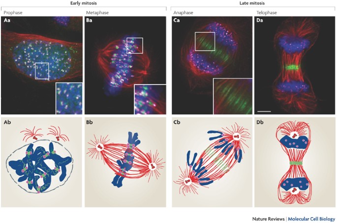
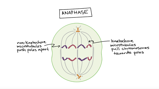
DOC Mitosis: Labeled Diagram Each chromosome has duplicated and now consists of two sister chromatids. At the end of prophase, the nuclear envelope breaks down into vesicles. Metaphase: The chromosomes align at the equitorial plate and are held in place by microtubules attached to the mitotic spindle and to part of the centromere. Anaphase: The centromeres divide.
Exam 1 Chapter 3 Flashcards | Quizlet Select all of the following events that occur during prophase. Multiple select question. The two centrosomes are replicated. The chromatids become condensed. The mitotic spindle begins to form. The nucleolus becomes less visible. The nuclear membrane begins to dissociate into small vesicles. meiosis. Eukaryotic cells that are diploid can divide by _____ to produce haploid sex …
Meiosis and Mendel’s Law of Segregation - STAR For simplicity, we are going to diagram only one chromosome, chromosome 1, and one hypothetical gene found on Chromosome 1, Gene 1, within a simplified version of a cell (only the cell membrane is shown). Gene 1 has two different variations, or alleles: A and a. Figure 1 illustrates what chromosome 1 looks like in a normal diploid cell: Figure 1 Following DNA …
The 4 Mitosis Phases: Prophase, Metaphase, Anaphase, Telophase Mitosis is important because it ensures that all new cells that are generated in a given organism will have the same number of chromosomes and genetic information. In order to accomplish this goal, mitosis occurs in four discrete, consistently consecutive phases: 1) prophase, 2) metaphase, 3) anaphase, and 4) telophase.
Cell division phases, types, and diagram - Jotscroll This diagram shows the cell division of prokaryotic cells. From the above image, the tangled line is the DNA and the remaining components are labeled as plasmids which are small rings of DNA that are also copied during binary fission and can be picked up in the environment from dead cells that disintegrate.
DOC Draw labeled diagrams showing the 10 stages of meiosis for ... 1. Draw labeled diagrams showing the 10 stages of meiosis for a diploid cell where 2N=6. Use 3 different shapes to represent the different chromosomes in each set, and use 2 different colors to represent the 2 sets of chromosomes. Make sure you show the number, shape, color and location of the chromosomes at each stage of division.
Prophase: Definition, Checkpoints, Diagram, and Examples Prophase: The prophase is marked by chromosomal condensation and disintegration of cellular components and assembly of cytoskeletons for cell division. RNA synthesis is inhibited. Metaphase: Nuclear membrane is eliminated completely chromosomes are completely condensed. The cytoskeleton - spindle fibers attach to the kinetochores. The chromosomes are aligned in the equatorial plate.
Chromosomes Fact Sheet - Genome.gov The unique structure of chromosomes keeps DNA tightly wrapped around spool-like proteins, called histones. Without such packaging, DNA molecules would be too long to fit inside cells. For example, if all of the DNA molecules in a single human cell were unwound from their histones and placed end-to-end, they would stretch 6 feet.
Meiosis I and Meiosis II; Life Cycles - Montgomery College Prophase II The chromosomes coil up, the nuclear membrane begins to disintegrate, and the centrosomes begin moving apart. 6. Metaphase II Spindle fibers form and sister chromatids align to the equator of the cell. ... Label the diagram (1-11) with phase or cell type (for what applies to the corresponding number), and chromosome number. ...
Bio Chapter 10 Flashcards - Quizlet Which event occurs only during prophase I of the first meiotic division? Synapsis of homologous chromosomes occurs. Which of the following is included in alternation of generations? All of the above : Zygote, spores, multicellular diploid stage (sporophyte), multicellular haploid stage (gametophyte) What is a locus? the precise location of a gene on a chromosome. How many …


(89).jpg)








Comments
Post a Comment